


 36 Min
 In Clinical reviews

Follow this topic

Bookmark

Record learning outcomes
|
||||||||||
Respiratory disease remains one of the leading causes of death in the UK and includes conditions such as lung cancer, COPD and asthma. Common pathological processes, including bronchoconstriction, inflammation and loss of lung elasticity, may present as breathlessness, wheeze, chest pain and cough.
However, non-respiratory conditions can also produce these symptoms, including gastrointestinal, cardiac and musculoskeletal causes. Diagnosis can therefore be challenging – particularly when respiratory symptoms coexist with other pathologies, especially CVD.
Cough is the most common respiratory symptom, although few conditions present with cough alone (see the summary table at the end of this article). Cough rarely indicates a need for urgent referral unless haemoptysis (coughing up blood) is present, whereas shortness of breath and chest pain are more commonly associated with serious pathology.
Establishing a diagnosis
A clear history of the presenting symptom or symptoms is central to diagnosis. As cough is the most common symptom associated with respiratory changes, as already mentioned, three questions – duration, nature and periodicity – should usually provide enough information to narrow the differential diagnosis to a small number of conditions.
For example, a four-month history of productive cough in a male that is worse in the morning would point towards COPD or bronchiectasis (see panels below).
| Cough questions | Implication (examples) |
| Duration | |
| Acute (<3 weeks) | Acute bronchitis Croup Infection Pulmonary embolism Pneumothorax |
| Sub-acute (3-8 weeks) | Upper airway cough syndrome (UACS) |
| Chronic (>8 weeks) | Asthma COPD Heart failure Lung abscess Malignancy |
| Nature | |
| Productive cough | COPD, cancer, heart failure, pneumonia |
| Non-productive | COVID-19, GORD, UACS, pneumothorax |
| Periodicity | |
| Worse in the morning: bronchiectasis, COPD Worse in the evening: croup, heart failure |
|
To differentiate between these conditions, ask specific questions about other manifestations of respiratory changes, including shortness of breath, wheeze and chest pain. After this targeted questioning, you are likely to have a high level of confidence in the diagnosis. At this point, a physical assessment may help confirm a clinical impression rather than establish the diagnosis.
| Four-month duration of cough symptoms suggests: | Four-month duration & productive cough: | Four-month duration, productive cough & worse in morning: |
| Asthma Bronchiectasis COPD GORD Heart failure Lung abscess Malignancy Nocardiosis Sarcoidosis TB Vocal cord dysfunction |
Bronchiectasis COPD Heart failure Lung abscess Malignancy Nocardiosis TB |
Bronchiectasis COPD |
Standard respiratory exam
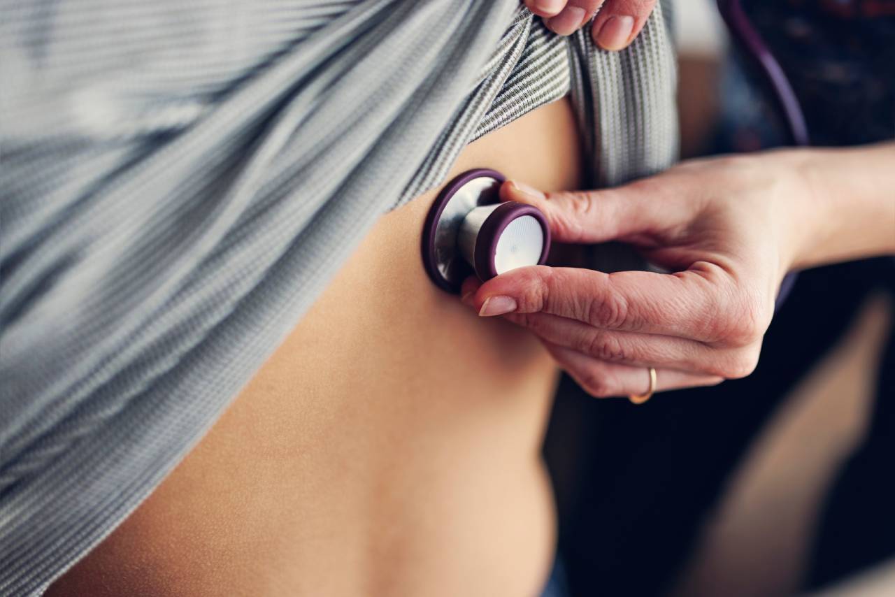
More than a third of pharmacists are now prescribers and this proportion is likely to rise substantially over the next few years. In principle, this means that respiratory physical examinations will fall within the scope of practice of many community pharmacists. The steps involved in a standard examination are summarised in Table 1.
In relation to a respiratory examination, Table 2 highlights the clinical implications of the various procedures, while Table 3 summarises the expected findings for respiratory conditions that may be encountered in community pharmacy.
Before conducting a respiratory examination in a community pharmacy setting, three questions should be considered:
1. Is it practical?
A standard respiratory examination usually takes between 8-10 minutes. Given current time pressures on pharmacists and pharmacy teams, finding this time may be challenging. A dedicated examination couch is also required, but this is not mandated for pharmacy consultation rooms. It is therefore unlikely that many current consultation rooms have either a couch or sufficient space to accommodate one.
| Table 1: Standard steps of a respiratory examination | |
| Steps | Observations |
| Introduction and explanation of procedure | Includes: wash hands; introduce self; outline process; gain consent; angle examination bed to 45°; adequately expose patient |
| General (‘end of bed’) inspection | Inspect for shortness of breath, cough, cyanosis, pallor, cachexia, scars |
| Pulse and respiratory rate | Pulse – assess rate and rhythm |
| Hands | Look for clubbing, peripheral cyanosis, asterixis, fine tremor |
| Face | Check for central cyanosis, oral candidiasis |
| Neck | Jugular venous pressure assessment, trachea deviation and assess cricosternal distance |
| Thorax | Chest shape and expansion |
| Palpate for apex beat and position | Check for displacement |
| Breath sounds | Percuss, auscultate and listen for vocal resonance |
| Palpate cervical lymph nodes | Check for enlargement |
| Oedema and DVT assessment | Look for ankle oedema and calf tenderness/pain |
| Summarise and close | Report back to patient and discuss next steps |
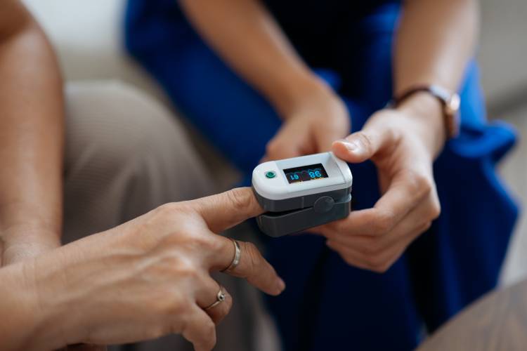
| The public is getting more familiar with pharmacists using diagnostic equipment like pulse oximeters |
2. Patient receptiveness
Public attitudes towards new pharmacy services, such as the New Medicine Service and Pharmacy First, are broadly positive. However, it is unclear whether this extends to activities that were until recently viewed as the remit of general practice.
Little research has been published on public receptiveness to pharmacists performing physical examinations. Although ear and throat observations are becoming more common through Pharmacy First, respiratory examinations are more involved and require the patient to be appropriately bare-chested. This might create a barrier to service expansion.
3. How useful is an examination?
Given that most diagnoses are made correctly through questioning alone, a full respiratory physical examination would appear to be of limited value in this context. Also, the most common conditions encountered in pharmacy, including viral upper respiratory tract infection, acute bronchitis and UACS, show similar physical findings and so are unlikely to be distinguished by full examination alone.

| A full respiratory physical examination may not always be necessary |
| Table 2: Findings and implications from a respiratory examination | |
| Finding | Implication |
| General inspection | |
| Cachexia | Cancer, COPD, heart failure |
| Pulse rate | Tachycardia seen in many respiratory conditions |
| Breathing rate | High = anxiety, acute asthma attack, pulmonary embolism, pneumothorax Low = heart failure, medication |
| Hands/nails | |
| Clubbing | Lung cancer, bronchiectasis, lung abscess |
| Peripheral cyanosis | Heart failure, asthma, pneumonia, croup |
| Flapping tremor – asterixis | Carbon dioxide retention suggests impaired ventilation attributable to COPD, asthma or bronchiectasis |
| Fine tremor | Beta-2 agonist use/overuse |
| Face | Conjunctival pallor (anaemia) Candida = steroid inhaler induced |
| Neck | |
| Raised jugular venous pressure | Heart failure, tricuspid regurgitation, constrictive pericarditis |
| Tracheal position | Deviation can indicate pneumothorax, tumour |
| Cricosternal distance | Reduced due to hyperinflation seen in asthma and COPD |
| Thorax | |
| Asymmetry of chest movement | Obstruction, infection, pneumothorax |
| Chest expansion | Lack of expansion can support a COPD diagnosis Asymmetry – pneumothorax, pneumonia |
| Apex beat and position | Ventricular hypertrophy, pneumothorax |
| Breath sounds | |
| Vocal resonance | Increased (suggests tissue density) – tumour, consolidation Decreased (suggests fluid or air) – pleural effusion, pneumothorax |
| Wheeze | Low pitched: COPD High pitched: asthma |
| Crackles | Fine: interstitial lung disease Medium: left ventricular failure or COPD Coarse: bronchiectasis |
| Pleural friction rub | Pleurisy, pneumonia or pulmonary embolism |
| Enlarged lymph nodes | Cancer, infection |
| Table 3: Physical examination – expected findings related to common and emergency respiratory conditions | ||||
| Condition | Inspection | Palpation | Percussion | Auscultation |
| Acute bronchitis | Normal or ↑ respiratory rate | Normal | Normal | Normal but added sounds of wheeze |
| Asthma | ↑ respiratory rate | ↓ chest wall movements | Normal or hyper-resonance bilaterally | Normal but added sounds of wheeze |
| COPD | ↑ respiratory rate | ↓ chest wall movements | Normal or hyper-resonance bilaterally | Normal but added sounds of wheeze or crackles |
| COVID-19 | Normal or ↑ respiratory rate | Normal or reduced expansion of affected side | Dull over affected side | Normal but added sounds of course crackles |
| PE* | ↑ respiratory rate | Normal | Normal | Normal |
| Pneumonia | ↑ respiratory rate | Normal or reduced expansion of affected side | Dull over affected side | Normal but added sounds of course crackles |
| Pneumothorax* | ↑ respiratory rate | Reduced expansion of affected side | Hyper-resonance on affected side | Absent breath sounds |
| UACS | Normal | Normal | Normal | Normal |
| Viral infection | Normal | Normal | Normal | Normal |
* Emergency situations
‘End-of-bed’ assessments
This type of general assessment can provide pharmacists with valuable additional information. For example:
- General inspection can help indicate how unwell the patient is and the severity of symptoms
- Respiratory rate – although often raised, it is usually normal in URTI, the most common condition likely to be encountered
- Oxygen saturation measured with an oximeter, which also estimates pulse rate, can raise suspicion of conditions like asthma, COPD and pneumonia
- Temperature measurement can be a useful indicator of infection
These assessments are quick and can be easily incorporated into a standard consultation. They may also help identify emergency situations, such as a respiratory rate of over 30 breaths per minute, pulse over 130 beats per minute, PO2 less than 92%, or using accessory muscles when breathing.
Management options
Depending on the diagnosis, this may include advice, self-care and pharmacological intervention. Common causes of cough are usually self-limiting.
Among acute causes of cough seen in community pharmacy, croup and pneumonia usually require prompt intervention and onward referral to a GP. For pharmacist prescribers, pharmacological intervention may be possible if within their clinical competence.
For croup, a single dose of oral dexamethasone (0.15mg/kg) should be given immediately.
For adults with suspected community-acquired pneumonia, a CRB65 score (0-4) should be calculated to assess mortality risk. Only those scoring zero should be considered for antibiotic treatment at home, with amoxicillin 500mg three times daily for five days.
In community pharmacy, patients may sometimes present with chronic cough without a formal diagnosis. Clinical signs and symptoms may suggest asthma or COPD, but objective testing is needed before treatment is initiated.
These tests are not routinely available in pharmacy, so referral to a GP is required. For example, suspected new asthma in patients aged over five years requires biochemical marker measurement or bronchodilator reversibility testing, while COPD requires spirometry for confirmation.
All patients presenting with chest pain and/or shortness of breath as the predominant symptom require onward referral, either to the GP or – if symptoms are severe – to A&E.
Summary: Conditions that present with respiratory symptoms
| Condition | Symptoms | ||||
| Shortness of breath | Wheeze | Chest pain | Cough | Other notable features | |
| Acute bronchitis | + | ++ | +++ | Fever, malaise | |
| Acute coronary syndrome (unstable angina and MI) | + | +++ | Nausea, sweating | ||
| Acute pulmonary oedema | +++ | + | Sweating, nausea | ||
| Anaemia | ++ | Fatigue, headache | |||
| Anxiety/panic attack | ++ | + (palpitation) | Light-headedness, sweating | ||
| Acute thoracic aortic dissection | +++ | Limb numbness | |||
| Asthma | ++ | ++ | + (tightness) | + | n/a |
| Biliary colic | +++ (epigastric) | Nausea & vomiting | |||
| Bronchiectasis | ++ | + | +++ | Fever, fatigue | |
| Cardiac arrhythmia | ++ | ++ | Dizziness | ||
| Cardiac tamponade | ++ | ++ | Rapid breathing, dizziness | ||
| Costochondritis | +++ | n/a | |||
| COPD | ++ | + | + | Fatigue | |
| Congestive heart failure | +++ | + | + | + | Fatigue, light-headedness |
| COVID-19 | + | +++ | Fever, loss of smell/taste | ||
| Croup | +++ | Onset in evenings | |||
| GORD | ++ (burning) | + | Taste disturbance | ||
| Lung abscess | ++ | ++ (pleuritic) | +++ | Fever, night sweats, weight loss | |
| Lung cancer | ++ | ++ | +++ | Fatigue, loss of appetite | |
| MI (Silent) | +++ | Malaise, nausea | |||
| Musculoskeletal pain | +++ | n/a | |||
| Nocardiosis | ++ (pleuritic) | +++ | Fever, night sweats, weight loss | ||
| Pulmonary embolism | +++ | ++ | + | Skin changes | |
| Peptic ulcer disease | +++ (epigastric) | n/a | |||
| Perforated oesophagus | +++ | Fever, vomiting, haematemesis | |||
| Pericarditis | + | +++ | + | Fever | |
| Pleurisy | + | +++ | + | n/a | |
| Pneumonia | ++ | + (pleuritic) | +++ | Fever, malaise | |
| Pneumothorax | ++ | ++ (pleuritic) | ++ | Signs of distress | |
| Psychogenic cough | +++ | n/a | |||
| Sarcoidosis | ++ | + | ++ | Fatigue, weight loss | |
| Sepsis | ++ | Fever, malaise | |||
| Stable angina | +++ | n/a | |||
| Thyroid disease | ++ | + (palpitation) | Temperature intolerance, GI disturbances | ||
| Tuberculosis | +++ | Fever, night sweats | |||
| Upper airway issue (aspiration, anaphylaxis or obstruction) | +++ | Signs of distress | |||
| Upper airway cough syndrome | +++ | Nasal congestion | |||
| Viral infection | +++ | Fever, cold symptoms | |||
| Vocal cord dysfunction | ++ | ++ | + | + | Stridor |
+ minor symptom; ++ common symptom; +++ major symptom
Red = requires urgent onward referral
| Useful resources | ||||||
|

Share:


In Clinical reviews